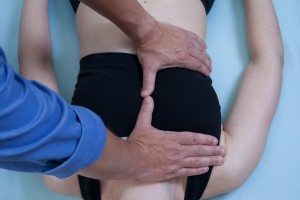
 Have you ever sat and wondered what happens behind that big bone in the front your chest? In a previous article, I briefly touched on the Thorax and its various articulations, common complaints in the area and some beginner assessment suggestions. This article is a brief discussion on the sternum and its interrelations/connections with the body.
My main goal for writing this article is to bring your awareness to an area often ignored by manual therapists for one reason or another. Treatment of this area can be truly amazing! Some of my most positive results for various injuries and pathologies have come from providing treatment to the tissues in this region.
Before I continue I must express that before attempting any work in this area, the Manual Therapist must have a working knowledge of the anatomy of the area. The books from Jean-Pierre Barral titled The Thorax & Visceral Manipulation are great starting points. The suggested treatments in these books will change how you normally assess & treat. If you have never treated a patient seated, you don’t know what you are missing.
I also suggest acquiring a Color Atlas of Anatomy. The images are some of the best I’ve seen nest to participating in an actual dissection class, which I feel should be part of all of our educations. It honestly is an invaluable experience.
I will not attempt to describe all the motions, movements, interconnections, relationships and so on that occurs with and around the sternum. There are volumes of books dedicated to this subject matter. Considering connections to start thinking about though, there are a few right under your perceptive little hands. We have tissue connections of between muscle, ligament, fascia, pleura, lung, neurovascular, lymphatic, osseous and so on. The sternum is also quite intimately connected not only to the thoracic spine but also to the cervical and lumbar spines and their respective fascial/connective tissues and vessels.
Fusion of the Sternebrae
Here are some interesting facts concerning when and which parts of the sternum fuse not only each other but with their friends the manubrium and xiphoid process.
The fusion of the different sternal elements takes place in relation to age, but is totally independent of the sex of the subject. The only differences for the most part are measurements of the sizes. The combined length of manubrium & body of sternum ranges from 101 mm to 192mm in male and 98 mm to 150mm in female.
Fusion between third and fourth sternebrae is usually complete by age 15. Fusion between second and third sternebrae was complete by age 21 and between first and second sternebrae the fusion was complete by age 21. Regarding fusion of xiphoid process with body of sternum it was complete by age 50. Fusion of manubrium with body of sternum starts at the age of 40 and it is complete by age 55. (Gautam, R. S., Shah, G. V., Jadav, H.R., Gohil, B.J.)
Connection examples range from Musculoskeletal (transversus thoracis – let’s see you “Pump Up” that one!), Osteoarticular (Intrasternal joints – didn’t see that one coming did you?), Myofascial (Endothoracic fascia to transverses thoracis), Visceralosseoss (Vertebropericardic Lig.), Musculovisceral (Phrenicopericardial Lig.), Intervisceral (Interpleural Lig.) and various other connections documented in the Barral books and various anatomy books.
As with all patients complaining of dysfunction in the thorax, a detailed thorough history must be done to rule out conditions that are outside our scope of practice and may require immediate emergent care. I have had a few times in my practice where I’ve caught a Myocardial Infarction and called 911. If this has yet to happen to you, be forewarned, it may. Most of the time though, the complaints are well within our scope.
Complaints
There are a number of patient complaints that we see every day that can be related to tissues here. Shortness of breath, tight exterior chest and or back, sharp specific and or shooting pain, deep pain, referred pain/tension, the inability to breathe deeply, indigestion burning within the cage with or without food, cervical tension radiating from the chest up to the cervical region, torsional strain feelings behind the sternum, pain over the heart where the heart has been ruled out, post surgical trauma, post MVA, strains, sprains, fractures, contusions, post impact injury of some sort, pain/tension within the chest sometimes described as being “in between”, respiratory challenges, pain around and or through the cage etc…
There are also complaints from respiratory conditions/pathologies such as asthma, scleroderma, emphysema, COPD, cystic fibrosis.
In cases such as these and many more, you must rely on your history taking skills, sensing/palpatory abilities and patient observational skills to make the determination of what a possible cause may be. Connections within the Thoracic cavity are not just limited to this area but are body wide and have great influence.
“The quieter the mind, the stiller the hands, the less movement we make, the more we are able to perceive involuntary movement.”
– James Jealous, DO
How to acquire information
In my previous article on the Thorax part 2, I offered suggestions on acquiring information from the tissues in this area. One way is to utilize standard orthopaedic testing and observational skills. These should become a regular part of your diagnostic assessment and can often lead you right to the source of the complaint.
Often I have found that a positional fault of one or more sternochondral ligamentous articular tissues/connections is the culprit of the discomfort. An investigation must start to determine why the tissues became dysfunctional. Here is where time is essential to go through the patients’ history. Sometimes the reason is obvious, others it may take a few weeks for a memory to surface.
Staying with this example, let’s discuss what can possibly happen to the surrounding areas, tissues and overall health of the patient if left untreated. Some patients ask if it will just get better and they can go on living their lives, or if it really needs to be treated since it doesn’t hurt any longer. In these cases we must remember the physiological process’s that are at work. Tissues go thru acute, sub-acute and chronic stages with various signs. Patients are mostly unaware of these processes. Now is the time to educate them as to what stage they are approximately in. Just because the site isn’t painful doesn’t imply that that the tissue(s) is/are functioning efficiently.
Thomas Schooley, DO wrote:
“It’s not perverted function but a wrong environment that results in the distorted appearance of function. Function is always true to its environment. Function is dependent upon its environment. Therefore, any change in any part of the environment that is not in tune or balance will apparently distort the function of the matter so involved.”
Assessing
Having progressed into a chronic stage of healing, why assess/treat the tissue? There are many things that can happen. An assessment of how the joint and surrounding tissues are moving, with respiration, with movement of the thorax, with movement of the extremities needs to be fully understood.
Are the Sternochondral joint tissues actually the primary dysfunctional tissues or are they a sign of some other dysfunctional tissues/process? What other tissues are being affected? Look at the sternochondral joints above & below; how are the intercostal spaces affected? What changes occur with inspiration and expiration? What changes occur with cervical rotation one way and the next? Note changes with cervical flexion and extension. Is there a corresponding dysfunction to the same level vertebralcostal joint?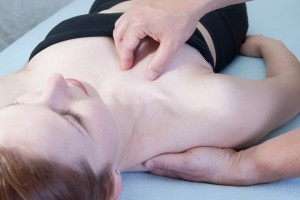
Concerning dysfunctions that occur on the left, I would refer you to “The Thorax” as it has great descriptions for the interconnections of the sternochondral, pericardial and costomediastinal tissues.
Avoid the RUT!
Don’t get into the rut of only assessing your patient in supine. Get them standing and seated. Incorporate active movements while you palpate the areas in concern. How do things change? Where is the pain/discomfort? How does it change with the different positions?
Hands on experience
One of the best educational experiences I’ve ever had was day 3-4 of a Gil Hedley Dissection course. “Fixed” forms, one that have been embalmed, obviously react differently than that of a live patient. The experiences gained with a cadaver are priceless and unforgettable. It will change your practice.
I was a TA for this course and was able to arrived early to get some “alone” time with the form we were exploring. With a Co-TA there to assist, I inserted a hand directly within the thoracic cage cavity and we began different compression techniques with rotation, torsions, and shears to attempt to fully understand what the tissues would be subjected to. It was amazing that even with my hand placed along the anterior vertebral bodies, I could feel the direct changes by having only a few ounces of influence placed from the anterior cage nearly 4 inches or more away.
We also noted the changes in directions of pull or directions of ease during these techniques by removing some adhesion’s/connections between tissues. I continually remember this experience every time I have a hand on a patient’s anterior thorax. You don’t have to use a lot of force to make a change in tissue. Less is often more in this area.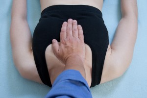
I challenge you to look at how the dysfunctional connections with-in the thoracic cage contribute to respiratory diaphragm dysfunctions, sinus congestion, cervicogenic headaches and complaints, iliopsoas dysfunctions leading to pelvic miss-alignments leading to postural and gait changes and much more.
Please remember that there are connections that are there by design and are necessary for life and general function, and then there are connections that are disrupting function of the vital organs and need to be attended to. Making the recognition of which to treat and which to leave is knowledge gained by education, study, experience, persistence, patients, focus, calmness and quietness.
Conclusion
As always, I hope that I have been able to serve you in some way with this information, be it a completely new perspective into your patients, be it some new thoughts about how to approach a treatment or provide you with a review and acknowledgement that what you are doing, as far as I’m concerned, is having an amazingly positive impact in improving the quality of life for your patients.
