Morphology of the Insertions of the Superficial Medial Collateral Ligament and Posterior Oblique Ligament Using 3-Dimensional Computed Tomography: A Cadaveric Study.
Abstract 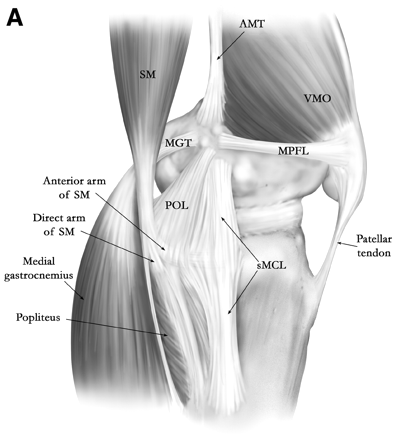
PURPOSE:
To describe the insertions of the superficial medial collateral ligament (sMCL) and posterior oblique ligament (POL) and their related osseous landmarks.
To describe the insertions of the superficial medial collateral ligament (sMCL) and posterior oblique ligament (POL) and their related osseous landmarks.
METHODS:
Insertions of the sMCL and POL were identified and marked in 22 unpaired human cadaveric knees. The surface area, location, positional relations, and morphology of the sMCL and POL insertions and related osseous structures were analyzed on 3-dimensional images.
Insertions of the sMCL and POL were identified and marked in 22 unpaired human cadaveric knees. The surface area, location, positional relations, and morphology of the sMCL and POL insertions and related osseous structures were analyzed on 3-dimensional images.
RESULTS:
The femoral insertion of the POL was located 18.3 mm distal to the apex of the adductor tubercle (AT). The femoral insertion of the sMCL was located 21.1 mm distal to the AT and 9.2 mm anterior to the POL. The angle between the femoral axis and femoral insertion of the sMCL was 18.6°, and that between the femoral axis and the POL insertion was 5.1°. The anterior portions of the distal fibers of the POL were attached to the fascia cruris and semimembranosus tendon, whereas the posterior fibers were attached to the posteromedial side of the tibia directly. The tibial insertion of the POL was located just proximal and medial to the superior edge of the semimembranosus groove. The tibial insertion of the sMCL was attached firmly and widely to the tibial crest. The mean linear distances between the tibial insertion of the POL or sMCL and joint line were 5.8 and 49.6 mm, respectively.
The femoral insertion of the POL was located 18.3 mm distal to the apex of the adductor tubercle (AT). The femoral insertion of the sMCL was located 21.1 mm distal to the AT and 9.2 mm anterior to the POL. The angle between the femoral axis and femoral insertion of the sMCL was 18.6°, and that between the femoral axis and the POL insertion was 5.1°. The anterior portions of the distal fibers of the POL were attached to the fascia cruris and semimembranosus tendon, whereas the posterior fibers were attached to the posteromedial side of the tibia directly. The tibial insertion of the POL was located just proximal and medial to the superior edge of the semimembranosus groove. The tibial insertion of the sMCL was attached firmly and widely to the tibial crest. The mean linear distances between the tibial insertion of the POL or sMCL and joint line were 5.8 and 49.6 mm, respectively.
CONCLUSIONS:
This study used 3-dimensional images to assess the insertions of the sMCL and POL and their related osseous landmarks. The AT was identified clearly as an osseous landmark of the femoral insertions of the sMCL and POL. The tibial crest and semimembranosus groove served as osseous landmarks of the tibial insertions of the sMCL and POL.
This study used 3-dimensional images to assess the insertions of the sMCL and POL and their related osseous landmarks. The AT was identified clearly as an osseous landmark of the femoral insertions of the sMCL and POL. The tibial crest and semimembranosus groove served as osseous landmarks of the tibial insertions of the sMCL and POL.
CLINICAL RELEVANCE:
By showing further details of the anatomy of the knee, the described findings can assist surgeons in anatomic reconstruction of the sMCL and POL.
By showing further details of the anatomy of the knee, the described findings can assist surgeons in anatomic reconstruction of the sMCL and POL.
Leave a Reply